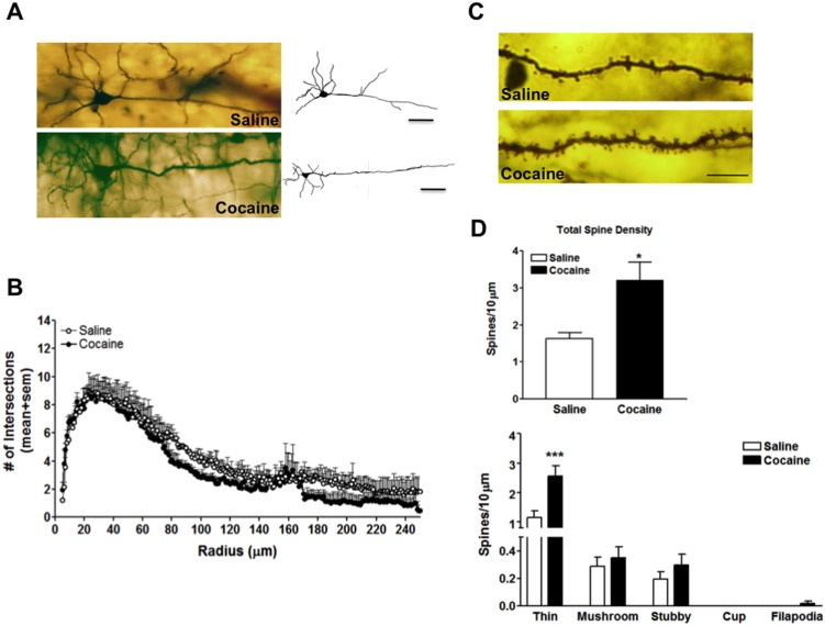
Figure 2. Single section Golgi analysis reveals alterations in dendritic branching and spine formation in the PFC following 7 days of abstinence from cocaine.
(A) Representative photomicrograph of a Golgi stain pyramidal neuron and tracing from yoked-saline control (top left, top right) and cocaine (bottom left, bottom right) treated rats scale bar = 50 µm (B) Sholl plot analysis of animals self-administering saline (open circles) and cocaine (black-filled circles) (C) Representative basal dendritic segment from a yoked-saline (top) and cocaine (bottom) neuron scale bar = 10 µm (D) Quantitative analysis of total spine number and spine density classified by spine sub-type from 4–5 segments from 5–7 neurons from each animal, n = 4 animals/group. *, p<0.05; ***, p<0.001.

