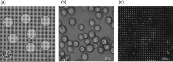
Figure 3.

(a) Schematic diagram and (b) microscope image of cells located on top of microwell array during transfection (c) lipoplexes containing FAM-ODN in the microwell array 4 h after transfection with A549 cells removed. Cell outlines are evident on the microwell array stamp following cell removal.
