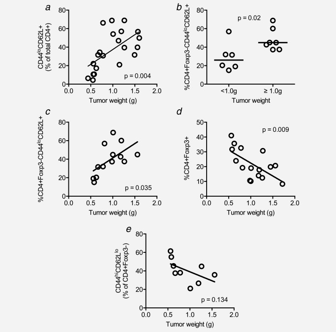
Figure 6.

The proportion of intratumoral naïve T cells increases significantly with tumor progression. Tumor cell suspensions were stained with CD4‐, CD44‐, CCR7‐ and CD62L‐specific mAbs and analyzed by flow cytometry. Proportions of CD4+CD44loCD62L+ cells were analyzed with respect to tumor weight at the time of sample preparation (a). Frequency of naïve CD4+ TILs in small versus large tumor (b). In (c), Foxp3+ cells were excluded from the analysis to more accurately demonstrate the relationship between naïve CD4+ cells and tumor weight. The relationship between CD4+Foxp3+ cells and tumor weight (d). The relationship between activated effector CD4+Foxp3− TILs and tumor weight (e).
