
Figure 3.
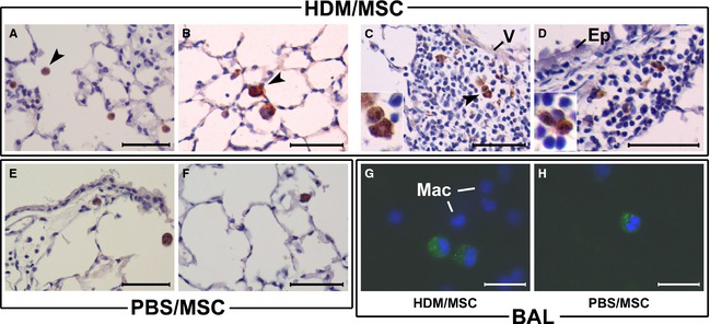
Location of mesenchymal stem cells (MSCs) in the lung. MSCs were retrovirally transduced to permanently express green fluorescent protein (GFP) and delivered intravenously (i.v.) to mice with established experimental asthma (house dust mite, HDM/MSC group, A–D, G) or disease control mice (phosphate‐buffered saline, PBS/MSC group, E–F, H). Upon 72 h (A) or 2 weeks (B–H) of continued HDM or PBS instillations following MSC infusion, GFP was detected by immunohistochemistry on lung sections (A–F; brown cytoplasmic signal) or from its native fluorescence on cytocentrifuged bronchoalveolar lavage (BAL) specimens (G, H). In the latter case, the GFP signal had a distinguishable granular pattern within the cytoplasm, whereas macrophage autofluorescence was below the photographing threshold (DAPI‐stained nuclei of surrounding monocyte/macrophages are shown for reference). GFP+ cells were found in the alveolar air spaces (A–B, E–F) and BAL of both the HDM/MSC and PBS/MSC mice, respectively, and also in the inflammatory infiltrates of HDM/MSC mice, in perivascular (C) and peribronchial (D) location. The GFP+ cells had a round‐shaped cell profile with a large cytoplasm/nucleus ratio and a cell size distinguishably larger than alveolar macrophages, all morphological characteristics consistent with cytological features of MSCs recruited to the lung; (C) and (D) insets show high‐magnification detail. V, vascular wall. Ep, airway epithelium. Mac, macrophages. Arrow heads signal examples of GFP+ MSCs. Scale bars: 100 μm in (A–F); 50 μm in (G, H).