
Figure 5. Expression of Shh, Gli1, S100A4 and E-cadherin in pancreatic cancer tissues analyzed by immunohistochemistry.
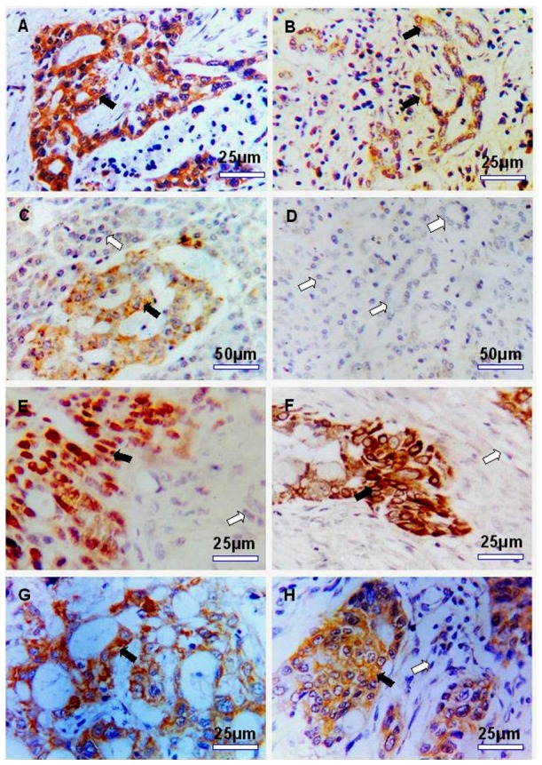
A: Strong cytoplasmic staining for Shh in pancreatic cancer cells (×400); B: Weakly positive staining for Shh in ductal complex of chronic pancreatitis tissues (×400); C: Weakly positive staining for Shh in islet cells of normal cancer side tissue (×200); D: Negtive staining for Shh in normal ductal epithelial/acinar cells of cancer side tissue (×200); E: Cancer cells show strong nuclear staining for Gli1 and normal cancer side tissues were negtive (×400); F: Strong positive perinuclear cytoplasmic and part nuclear staining for Gli1 in pancreatic cancer cells (×400); G: Strong positive cytoplasmic staining for S100A4 in pancreatic cancer cells (×400); H: Positive cytoplasmic staining for E-cadherin in pancreatic cancer cells. Black arrows point to positive staining area and white arrows point to negtive staining area.