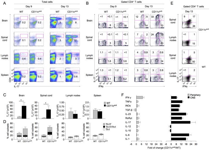
Figure 2. Potent Th17 differentiation uncovered in the inflamed CNS of CD11cdnR mice.
(A–D) Flow cytometry of CD4 versus IL-17 among total cells (A) and IL-17 versus IFNγ among gated CD4+ T cells (B) in brain, spinal cord, lymph nodes, and spleen of CD11cdnR (n = 8) and wild-type (WT) (n = 8) mice at days 9 and 13 post-immunization. (C) Numbers of Th17 (CD4+IL-17+) cells in brain, spinal cord, lymph nodes, and spleen of CD11cdnR (black) versus wild-type (WT) (gray) mice at the peak of EAE (day 13). (D) Percentages of Th17 (IL-17+IFNγ−) (gray), Th1 (IL-17−IFNγ+) (white), and Th1/Th17 (IL-17+IFNγ+) (black) cells in brain, spinal cord, lymph nodes, and spleen of CD11cdnR versus wild-type (WT) mice at the peak of EAE (day 13). (E) Spinal cord and draining lymph nodes were isolated from CD11cdnR (n = 4) and wild-type (WT) (n = 4) mice on day 13 post-immunization and total mononuclear cells were cultured in the presence of 50 µg/ml MOG peptide for 24 hours. Plots show the distribution of IL-17 versus IFNγ among gated CD4+ T cells in response to MOG re-stimulation from spinal cord versus draining lymph nodes. (F) SYBR Green quantitative PCR of the indicated genes in the CNS (black) and periphery (gray) of CD11cdnR (n = 6) and wild-type (n = 6) mice at the peak of EAE (day 13). Data were analyzed using the 2−ΔΔCt (cycle threshold) method, and results are expressed as the fold of change in CD11cdnR versus wild-type organs. Data are representative of three (A–D) and two (E–F) independent experiments. Mean ± s.e.m. (C–D). *P<0.05 (C).

