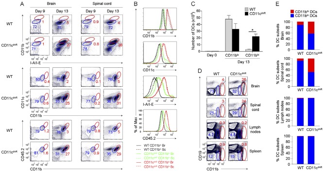
Figure 3. Inflamed CNS of CD11cdnR mice revealed massive production of highly mature DCs lacking TGF-βR signaling.
(A) Flow cytometry of CD11b versus I-A/I-E, CD11c versus CD11b, and CD45.2 versus CD11b in the brain and spinal cord of CD11cdnR and wild-type (WT) mice at days 9 (n = 5) and 13 (n = 8) post-immunization. Plots are from mononuclear cells. Gates delineate two myeloid cell subsets expressing high (red) and low (blue) levels of DC maturation markers, and numbers represent the percentages of cells in red gates. (B) Overlay of gated CD11blo (black and green) and CD11bhi (red) cell subsets from the brain (Br; solid) and spinal cord (Sc; dashed) of CD11cdnR (green and red) and wild-type (WT) (black) mice at peak of EAE (day 13). Histograms display the fluorescence intensity of surface expression of CD11b, CD11c, I-A/I-E, and CD45.2. (C) Numbers of DC subsets (CD11blo and CD11bhi) recovered from the CNS of CD11cdnR (black) and wild-type (WT) (gray) mice at peak of EAE (day 13). (D) Flow cytometry of CD19 versus CD11b in brain, spinal cord, lymph nodes, and spleen of CD11cdnR and wild-type (WT) mice at peak of EAE (day 13). (E) Ratio of CD11blo (blue) versus CD11bhi (red) DC subsets in brain, spinal cord, lymph nodes, and spleen of CD11cdnR and wild-type (WT) mice at peak of EAE (day 13). Data are representative of three independent experiments. Mean ± s.e.m. (C). *P<0.05 (C).

