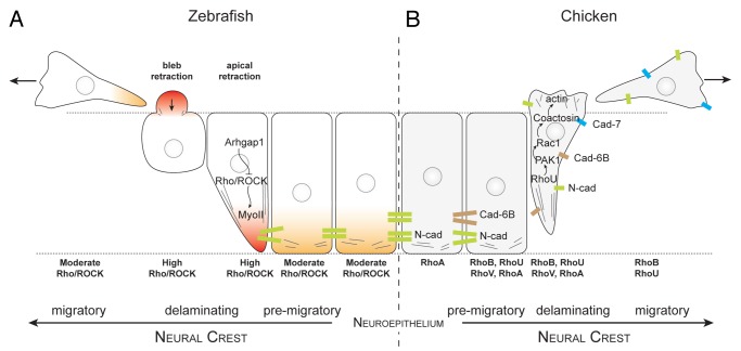
Figure 3. Rho signaling during neural crest delamination in zebrafish and chick embryos. (A) Current view of zebrafish neural crest delamination. Arhgap1 restricts Rho activity to the apical side. NC cells exit the neural tube by retracting their apical side and forming blebs on their basal side. (B) Current view of chick neural crest delamination. Changes in cell-cell adhesion repertoire occur concomitantly with changes in cell contractility. NC cells may exit by first disrupting their cell-cell adhesion and then acquire motility. Alternatively, they may force their way out by forming protrusions and mechanically disrupting their junction with other neuroepithelial cells by pulling the cell body. Delaminating chick NC cells express RhoA, B, U and V. The respective roles of the Rho proteins are open to conjecture although it is likely that RhoU controls protrusion formation via PAK1/Rac1 whereas RhoB appears to be involved in cell-cell adhesion disassembly.

An official website of the United States government
Here's how you know
Official websites use .gov
A
.gov website belongs to an official
government organization in the United States.
Secure .gov websites use HTTPS
A lock (
) or https:// means you've safely
connected to the .gov website. Share sensitive
information only on official, secure websites.
