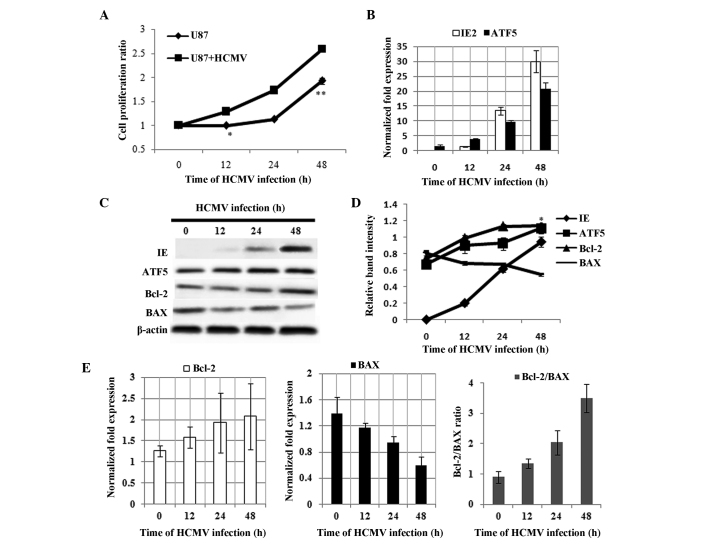
Figure 1.
Effect of HCMV infection on cell proliferation and expression of ATF5, Bcl-2 and BAX in U87 cells. (A) Cell proliferation was measured using a 3-(4,5-dimethylthiazol-2-yl)-2,5-diphenyltetrazolium bromide assay. Data are presented as the mean ± SD of three independent experiments (*P<0.05 and **P<0.01 vs. U87 + HCMV). (B) qPCR analyses showing the expression levels of IE2 and ATF5 mRNA in U87 cells following HCMV infection for 0, 12, 24 and 48 h. (C) Western blot analysis of IE genes, ATF5, Bcl-2 and BAX protein in HCMV-infected U87 cells. (D) Relative expression of IE, ATF5, Bcl-2 and BAX, all vs. β-actin in U87 cells infected with HCMV for 0, 12, 24 and 48 h, according to the results of figure 1C. Data are presented as the mean ± SD (*P<0.01 vs. 0 h ATF5 expression). (E) qPCR analyses of Bcl-2 and BAX mRNA expression in U87 cells following HCMV infection, where the ratio of Bcl-2/BAX was calculated. HMCV, human cytomegalovirus; ATF5, activating transcription factor 5; Bcl-2, B-cell lymphoma/leukmia-2; BAX, Bcl-2-associated X protein; SD, standard deviation; qPCR, quantitative polymerase chain reaction; IE, immediate-early.

