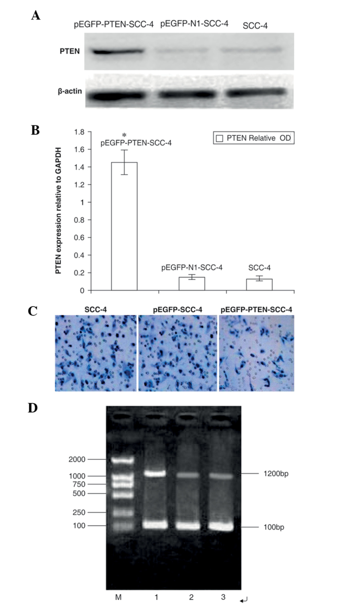
Figure 2.

Effects of PTEN vector transfection on PTEN expression in SCC-4 cells. (A) Untransfected cells (lane 1) and cells transfected with empty vector (lane 2) or PTEN (lane 3) were subjected to western blotting using anti-PTEN antibodies. β-Actin was used as a loading control. (B) Quantification of western blots to measure PTEN protein expression. The OD of PTEN (relative to GAPDH) was as follows: 1.45±0.14, 0.17±0.02 and 0.15±0.03 in the pEGFP-PTEN-SCC-4, pEGFP-N1-SCC-4 and SCC-4 groups, respectively. (C) Effects of PTEN expression on the invasion of SCC-4 cells. Untransfected SCC-4 cells and cells transfected with pEGFP-SCC-4 or pEGPF-PTEN-SCC-4 were subjected to Transwell invasion assays. The number of cells with penetrated membranes in the pEGPF-PTEN-SCC-4 group was evidently less than in the SCC-4 and pEGFP-SCC-4 groups. (D) Reverse transcription-polymerase chain reaction for PTEN gene mRNA expression. M, DNA maker; 1, pEGFP-PTEN-SCC-4; 2, pEGFP-N1-SCC-4; and 3, SCC-4. PTEN, phosphatase and tensin homolog; pEGFP, phosphorylated enhanced green fluorescent protein; SCC, squamous cell carcinoma; OD, optical density.
