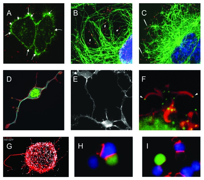
Figure 4. Illustrations of viral use and manipulation of the cytoskeleton through Rho GTPase signaling during virus entry, egress, and interaction with immune cells. (A) Actin(green)-dependent surfing of Avian Leukosis Virus particles (red) along filopodia to the cell body during virus entry. (B-C) Rac1-mediated microtubule(green)-based transport of adenovirus particles (red) to the nucleus (blue) during virus entry. Cells in (C) were treated with the Rac1 inhibitor NSC23766 and show reduced association of virus particles with microtubules and reduced accumulation of virus particles at the nuclear rim. (D-E) Similar phenotype of actin-based cell projections induced by the alphaherpesvirus pseudorabies virus US3 protein kinase (D) and vaccinia virus F11 protein (E). (D) shows actin (red), microtubules (blue) and virus particles (green), (E) shows actin cytoskeleton. (F) Actin(red)-dependent expulsion of vaccinia virus particles (green) from infected cells during virus egress. (G-I) Effects of HIV Nef-PAK2 signaling on HIV interaction with immune cells. (G) Nef-mediated production of virus particle (white)-tipped filopodia (red) in dendritic cells. (H-I) HIV (H) is unable to disturb actin-dependent immunological synapse (red) formation between infected Jurkat T cells (green) and Raji B cells (blue), whereas wild-type HIV disturbs immunological synapse formation (I). Image sources: (A) Lehmann et al., 2005, J Cell Biol, (B,C) Warren and Cassimeris, 2007, Cell Mot Cytoskeleton, (D) Image courtesy of Herman Favoreel, (E) Valderrama et al., 2006, Science, (F) Cudmore et al., 1995, Nature, (G) Aggarwal et al., 2012, PLoS Path, (H, I) Rudolph et al., 2009, J Virol.

An official website of the United States government
Here's how you know
Official websites use .gov
A
.gov website belongs to an official
government organization in the United States.
Secure .gov websites use HTTPS
A lock (
) or https:// means you've safely
connected to the .gov website. Share sensitive
information only on official, secure websites.
