Figure 1.

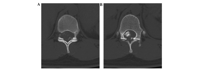
Axial computed tomography scan slice at thoracic level 11, showing extension of the neural foramina on the right, with (A) foraminal enlargement and (B) an isodense mass with calcification.

Axial computed tomography scan slice at thoracic level 11, showing extension of the neural foramina on the right, with (A) foraminal enlargement and (B) an isodense mass with calcification.