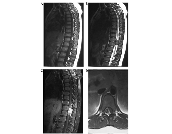
Figure 2.
Pre-operative magnetic resonance imaging showing a mass with iso- to hypointensity on the (A) T1-weighted image and (B) mixed hypointensity on the T2-weighted image. (C) Heterogeneous enhancement on the T1-weighted image with gadolinium. (D) The spinal cord was severely compressed and displaced to the left.

