
Figure 3.
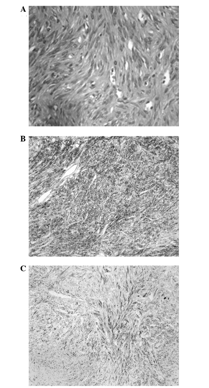
Photomicrographs of the surgical specimens illustrating (A) spindle-shaped cells arranged in fascicles with blunt-ended nuclei (hematoxylin and eosin staining; original magnification, ×400), (B) strong positivity for smooth muscle actin (immunohistochemical staining; original magnification, ×100) and (C) strong positivity for desmin (immunohistochemical staining; original magnification, ×100).