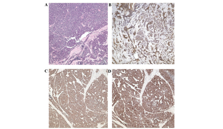
Figure 2.
(A) Microscopically, the lesions were composed of sheets of small atypical cells with little cytoplasm and large, round nuclei (stain, hematoxylin and eosin; magnification, ×100). Immunohistochemical EnVision™ staining revealed that the neoplastic cells were positive for (B) cytokeratins, (C) synaptophysin and (D) chromogranin A (magnification, ×100).

