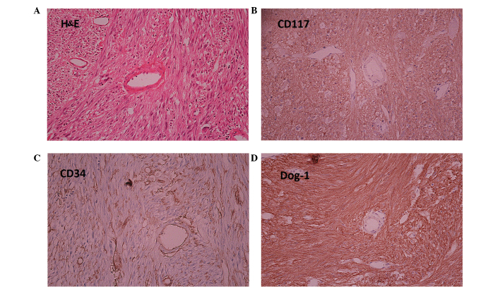
Figure 4.
H&E and immunohistochemical staining of gastrointestinal stromal tumors (magnification, ×400). (A) H&E staining demonstrated a different cellular morphology compared with the adenocarcinoma as it was spindle-shaped. Immunohistochemical analysis showed positive staining for (B) CD117, (C) CD34 and (D) Dog-1. H&E, Hematoxylin and eosin; CD, cluster of differentiation; Dog-1, discovered on GIST-1.

