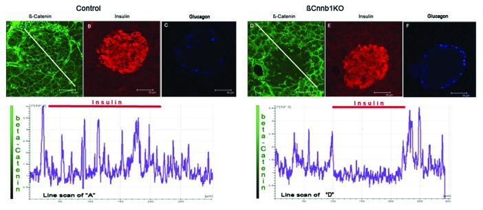
Figure 5. Deleting of Ctnnb1 gene in pancreatic β-cells (βCtnnb1KO) leads to elimination of β-catenin from insulin positive cells in islets of Langerhans. Pancreatic sections stained for - β-catenin (GREEN; A, D), insulin (RED; B, E) and glucagon (BLUE, C, F). Mouse samples: (A, B, C) control (Ctnnb1fl/fl[Tmx]); (D, E, F) double-transgenic Ctnnb1KO, (Ctnnb1fl/fl:MIP1-CreERT[Tmx]). Note the decreased β-catenin staining in the islet area of Ctnnb1KO pancreas despite the preserved β–catenin staining of surrounding acinar tissue. The graph under the images represents intensity of staining along the white line shown in the β-catenin-stained panels. Note the decreased level of β-catenin intensity under the area of insulin staining indicated by red bars.

An official website of the United States government
Here's how you know
Official websites use .gov
A
.gov website belongs to an official
government organization in the United States.
Secure .gov websites use HTTPS
A lock (
) or https:// means you've safely
connected to the .gov website. Share sensitive
information only on official, secure websites.
