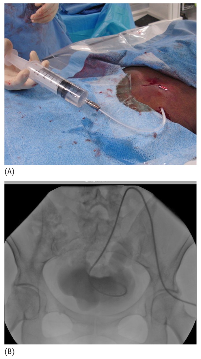
Figure 15 —

(A) Photographic and (B) fluoroscopic images show nonionic contrast injected through the catheter under fluoroscopic visualization to exclude any catheter kinking in the subcutaneous tunnel or peritoneal entry site, and to confirm the proper location of the curled distal tip within the pelvic portion of the peritoneal cavity.
