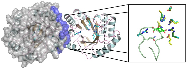
Figure 5. Crystal structure of efDHQase.
The biological assembly of efDHQase is a homodimer. Alpha-helices, β-strands, and loops are shown as cartoon representations and colored light blue, golden, and pink, respectively, to visualize the (β/α)8-barrel scaffold on each monomer. Residues at the dimer interface are highlighted purple on the surface (grey) of one monomer. Residues involved in ligand binding are shown as sticks and colored cyan. Comparison of efDHQase's active center with homologous 3-dehydroquinate dehydratases from Salmonella enterica (seDHQase) and Clostridium difficile (cdDHQase) is shown in the magnified view on the right. The structures were superimposed onto each other using the Cα atoms of the ligand-binding residues, with the residues from seDHQase and cdDHQase colored dark green and yellow, respectively. The pre-dehydration intermediate covalently-attached to seDHQase is drawn as green sticks. Part of the β8-α8 loop from seDHQase is shown as light green cartoon representations.

