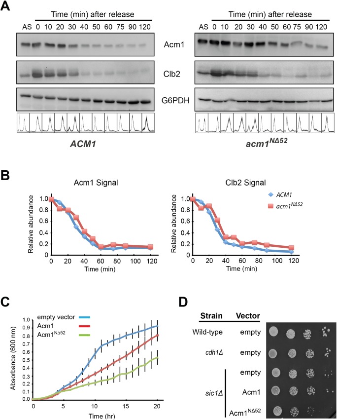
Figure 7. Acm1NΔ52 is still cleared at mitotic exit, but constitutive expression impairs growth of sic1Δ cells.
A) YKA859 cells carrying either pHLP117 (for expression of 3HA-Acm1) or pHLP505 (for expression of Acm1NΔ52-ProtA) were arrested at metaphase by methionine repression of PMET3-CDC20 and then released in the absence of methionine. Cells were collected at regular intervals and analyzed by immunoblotting using antibodies against Acm1, Clb2, or G6PD (loading control). B) Quantitation of chemiluminescent immunoblots from panel A. Data are the average of 4 independent experiments. C) Growth of sic1Δ cells transformed with either an empty vector, pHLP361 (PADH-Acm1-ProtA) or pHLP363 (PADH-acm1NΔ52-ProtA) were compared using a plate reader to measure absorbance at 600 nm. Data are the average of three independent experiments with standard deviation error bars. D) Serial 10-fold dilutions of the strains from panel C as well as isogenic wild-type and cdh1Δ strains were spotted and grown on selective agar plates.

