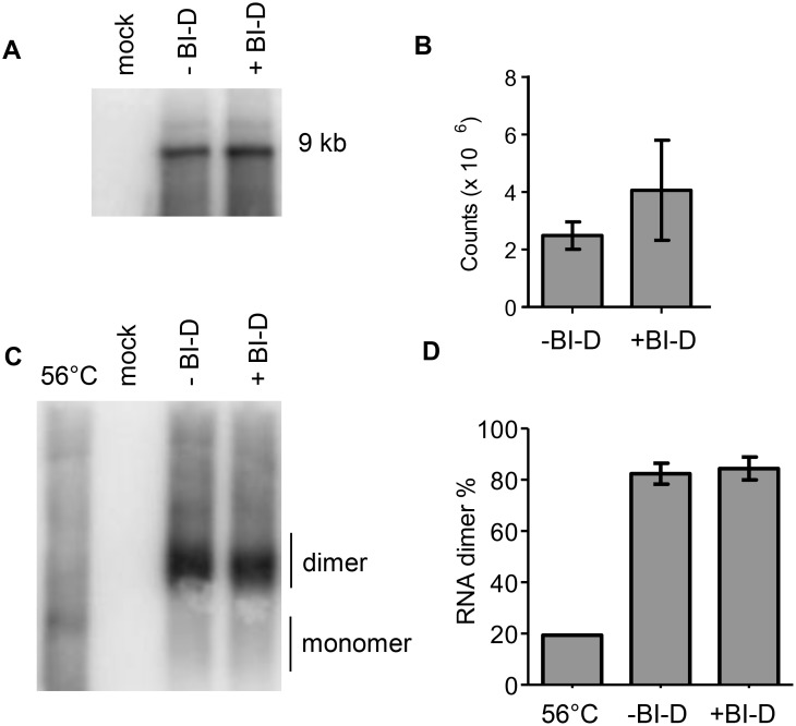
Figure 2. HIV-1 RNA packaging and dimerization.
Viral RNA was isolated from virion particles produced in the presence or absence of BI-D. For mock samples, RNA was isolated from pBluescript-SK+ transfected cells. A. Virus RNA was analyzed on a denaturing northern blot to measure RNA packaging. The position of the full-length 9 kb viral genome is indicated. B. Quantification of the 9-kb viral RNA detected in panel A. The average value (N = 3) with SD is shown. C. Viral RNA was analyzed on a non-denaturing northern blot to analyze the dimerization status. The position of the dimer and monomer are indicated. To identify the position of the monomer, the – BI-D RNA sample was incubated at 56°C for 10 min before analysis. D. The monomer and dimer bands observed in panel C were quantified to measure the level of dimerization. The average value with SD is shown (N = 3).

