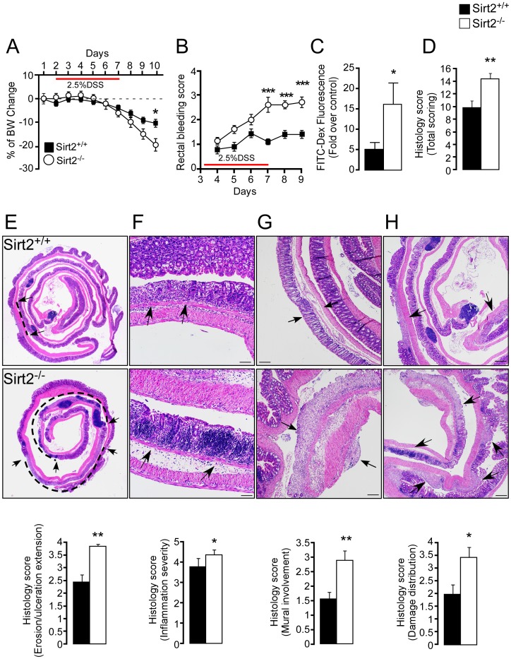
Figure 3. Sirt2−/− mice are more sensitive to DSS-induced colitis compared to Sirt2+/+ animals.
(A–D) Severity of DSS-induced colitis was determined by body weight change (A), rectal bleeding scores (B), intestinal permeability (C), and histological scores (D) in Sirt2+/+ and Sirt2−/− mice. n = 10/group. (E–H) Histological changes in the intestine of the DSS-treated Sirt2+/+ and Sirt2−/− mice. Representative images demonstrating the extension of erosion/ulceration (arrows and dashed line indicate the extent of ulceration) (E), inflammation severity (arrows indicate leukocytic infiltrate and follicular aggregates) (F), mural involvement (arrows indicate transmural infiltration) (G), and damage distribution (arrows indicate sites of damage) (H). Corresponding histological scores are shown (lower panels). n = 10/group. Scale bar = 20 µm. Results are expressed as the mean ± SEM. *P<0.05; **P<0.01; ***P<0.001.

