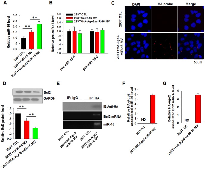
Figure 3. HA-Ago2 in MVs guides the function of secreted miR-16 in recipient cells.
A and B, The expression levels of miR-16 (A) and pre-miR-16 (B) in 293T cells detected using RT-qPCR. 293T cells were incubated with MVs secreted from HeLa cells transfected with random oligonucleotides (CTL), miR-16 (miR-16) or co-transfected with the miR-16 mimic and HA-Ago2 plasmid (HA-Ago2/miR-16). C, Immunofluorescence staining of HA-Ago2 in 293T cells with anti-HA antibody. D, The protein levels of Bcl2 in 293T cells detected using Western blot analysis. E–G, The direct association of miR-16 (E and F) and Bcl2 mRNA (E and G) with HA-Ago2. HA-Ago2 was immunoprecipitated from recipient 293T cell lysate using anti-HA antibody, and the levels of miR-16 (F) and Bcl2 mRNA (G) associated with HA-Ago2 or control IgG were detected using RT-qPCR. **, P<0.01.

