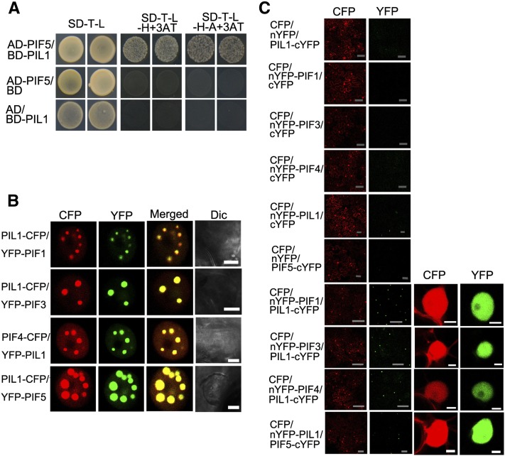
Figure 7.
PIL1 Physically Interacts with PIFs.
(A) Yeast two-hybrid assay of the PIL1–PIF5 interaction. Yeast cells transformed with the indicated combinations of constructs were grown in nonselective (SD-T-L) or selective media with 2 mM 3AT (SD-T-L-H+3AT or SD-T-L-H-A+3AT).
(B) PIL1 and PIFs (PIF1, PIF3, PIF4, and PIF5) colocalize to NBs in tobacco cells. Dic, differential interference contrast. Bars = 5 μm.
(C) BiFC assay of the PIL1–PIFs (PIF1, PIF3, PIF4, and PIF5) interactions in tobacco leaf cells. CFP serves as an internal control. The left two panels show the wide field view to indicate the BiFC efficiency. The right two panels show a single nucleus at high magnification. Gray bars = 100 μm; white bars = 5 μm.

