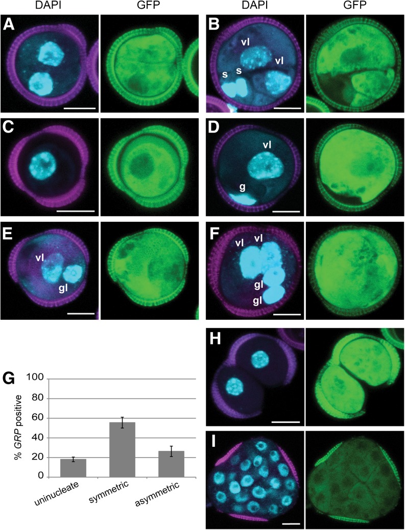
Figure 2.
Embryo Identity in Microspore Culture Is Not Dependent on Cell Division or Division Symmetry.
(A) to (F) proGRP:GUS-GFP embryo marker expression after 3 d of culture. DAPI staining (blue), autofluorescence (magenta), and GFP expression (green).
(A) Symmetrically divided microspore showing GRP expression in both cells.
(B) GRP expression in the two symmetrically divided vegetative-like cells, but not the smaller generative/sperm-like cells.
(C) GRP-positive microspore
(D) GRP expression in the vegetative-like cell, but not in the smaller generative-like cell.
(E) and (F) GRP expression in both the vegetative- and generative-like cells. Note that the generative-like nuclei in (E) and (F) are larger and the DNA is less condensed than in (D).
(E) Asymmetrically divided bicellular structure.
(F) Multinucleate structures.
(G) Proportion of microspores and symmetrically and asymmetrically divided structures that express proGRP:GFP-GUS at day 3 of culture. Structures where GRP was only expressed in the vegetative cell of binucleate (D) were not included in the graph.
(H) and (I) GRP-positive structures found at 6 d of culture.
(H) Sporophytic structure prematurely released from the exine.
(I) Proembryo enclosed in the exine. Note that GFP fluorescence was weaker in multinucleate proembryos than in few celled embryos ([A] to [F]).
Error bars in (G) indicate the se of three replicates. vl, vegetative-like nucleus; gl, generative-like nucleus. Bars = 10 μm.

