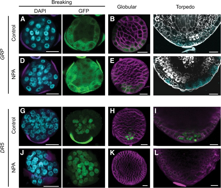
Figure 6.
Auxin Polar Transport Is Not Required for Microspore Embryo Polarization.
(A) to (F) Establishment of the embryo basal domain, marked by proGRP:GFP-GUS.
(G) to (L) Localization of auxin response maxima by proDR5:GFP expression.
Comparison of control and NPA-treated embryos at the stage where the exine starts to break ([A], [D], [G], and [J]), at the globular stage after release from the exine ([B], [E], [H], and [K]), and in the root pole at the torpedo stage at the torpedo stage ([C], [F], [I], and [L]). DAPI staining of the nuclei (blue) and GFP expression (green) are shown separately. FM4-64 (magenta) was used to stain membranes in globular stage embryos. GUS staining of proGRP:GFP-GUS lines (light blue) was combined with starch and cell wall localization by Pseudo-Schiff-propidium iodide staining (white). Bars = 20 μm.

