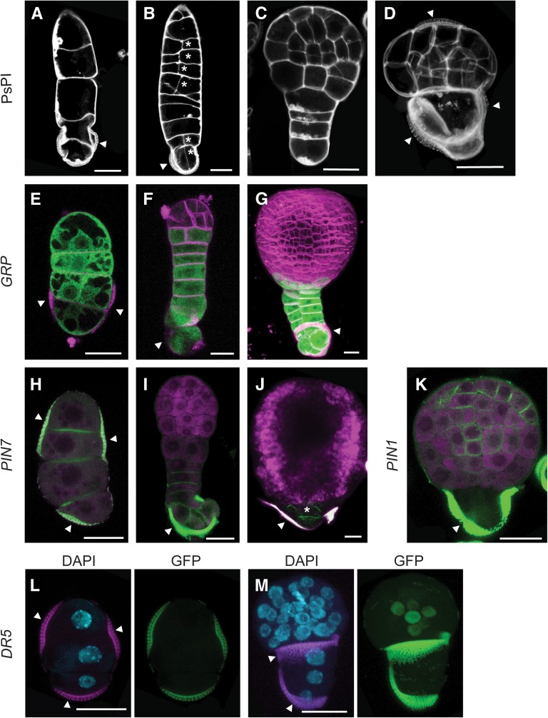
Figure 7.
Domain Specification in Suspensor-Bearing Microspore Embryos Is Similar to Zygotic Embryos.
(A) to (D) Suspensor morphologies observed in microspore culture.
(A) Uniseriate suspensor.
(B) Suspensor with aberrant longitudinal divisions (asterisk).
(C) Microspore embryo with suspensor and embryo proper.
(D) Microspore embryo with two domains, enclosed in the exine. Arrowheads point to the three exine locules enclosing the proembryo.
(E) to (G) proGRP:GFP-GUS expression. GFP is shown in green and FM4-64–stained membranes in magenta.
(E) Uniseriate suspensor.
(F) Suspensor-bearing embryo at the early globular stage.
(G) Transition-stage suspensor-bearing embryo, with two cell files.
(H) to (J) proPIN7:PIN7-GFP expression (green) in the suspensor. Propidium iodide stain or autofluorescence (magenta)
(H) Uniseriate suspensor.
(I) Globular stage embryo with suspensor
(J) Transition stage embryo with a rudimentary suspensor.
(K) proPIN1:PIN1-GFP expression (green) in a globular stage suspensor-bearing embryo.
(L) and (M) proDR5:GFP expression (green) in the embryo proper. DAPI nuclear stain (blue) and autofluorescence (magenta).
(L) DR5 is not expressed in the suspensor.
(M) Suspensor-bearing embryo at the globular stage showing DR5 expression in the inner cells at the basal region of the embryo proper.
Arrowhead, exine. Bars = 20 μm.

