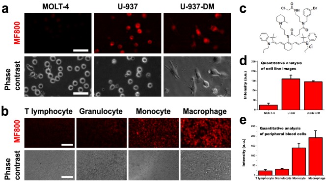
Figure 1. Selectivity of MF800 for monocytes/macrophages in a live-cell labeling assay.
(a) Labeling of MF800 to MOLT-4 (lymphocyte) and U-937 (monocyte) cell lines, and in U-937-derived macrophages (U-937-DM) after incubation with 2 µM of the probe for 1 hour at 37°C. All NIRF images had identical exposure times and were normalized to the peak signal. Scale bars are 50 µM. (b) Probing human leukocytes collected from erythrocyte-lysed fresh blood and selected by magnetic separation. Images were obtained with the identical measurement and analysis conditions for NIRF after incubation with 6 µM of MF800 for 1 hour at 37°C. Scale bars are 100 µM. (c) Chemical structure of MF800. Image-based NIRF quantification from (d) the MF800 stained cell lines (n = 3, three different experiments) and (e) peripheral blood cells (n = 3, three different donor samples). Data are presented as mean ± s.d. a.u., arbitrary units.

