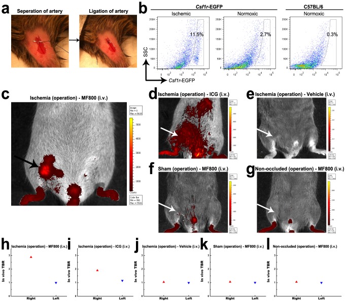
Figure 3. In vivo NIRF imaging of hind-limb ischemia with MF800.
(a) To generate hind-limb ischemia, an incision was made in the skin, surgical thread was inserted underneath the femoral artery (left); then the right femoral artery was ligated by a triple surgical knot (right). (b) Flow cytometry analysis of digested tissues 3 days after ligation shows a higher proportion of csf1r-EGFP-positive macrophages in the ischemic right hind-limb (11.5 %, left) compared with the normoxic left hind-limb (2.7 %, middle) in csf1r-EGFP transgenic mice. Cells from the normoxic hind-limbs of naïve C57BL/6 mice were used for control (0.3 %, right). (c–l) in vivo NIRF imaging and signal quantifications of mouse models of hind-limb ischemia receiving (c, h) MF800 (n = 4), (d, i) ICG (n = 2) or (e, j) vehicle control (n = 2) intravenously. For negative controls, (f, k) sham-operated (n = 2) and (g, l) non-occluded (n = 2) groups were imaged after i.v. injection of MF800. Pictures shown are merges of pseudocolored fluorescence and white light images with corresponding color lookup tables 4 hours after i.v. injection. MF800 illuminated ischemic regions with significant macrophage recruitment (arrow) whereas ICGs were deposited nonspecifically. The TBR value for the ischemic right hind-limb in the MF800-injected mice was higher than that of the contralateral paw, as well as ICG-injected and other control groups.

