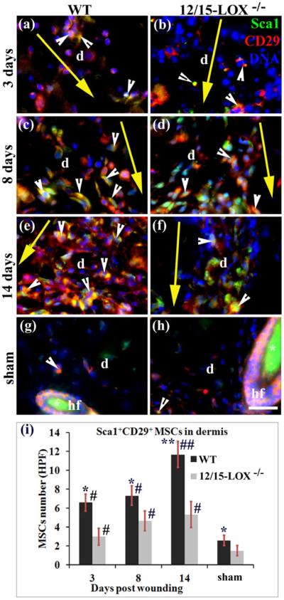
Fig. 2. 12/15-LOX deficiency reduces densities of Sca1+CD29+ MSCs in the dermis of wounded skin.
Images in (a) and (b), images in (c) and (d), and images in (e) and (f), are for 3, 8 and 14 dpw, respectively. Images in (g) and (h) are for sham (unwounded skin). Images in (a), (c), (e) and (g), and images in (b), (d), (f) and (h), are for C57BL/6 (WT) and 12/15-LOX−/− mice, respectively. Immunofluorescent co-staining and microscope imaging were conducted on cryosections of each wound with a 10 mm margin of surrounding skin, or skin at the equivalent location of sham. (i) There are more cells co-stained (yellow) for Sca1 and CD29 (i.e, Sca1+CD29+ MSCs) in the wounded dermis of WT mice compared to 12/15-LOX−/− mice for 3, 8 and 14 dpw. Each value is the average number per HPF of Sca1+CD29+ MSCs from six mice per group ± SD. *p < 0.05 and **p < 0.01 are for WT versus 12/15-LOX−/−; #p < 0.05 and ##p < 0.01 are for the wounded versus sham of the same type of mice. HPF, high power field (63X); d, dermis; hf, hair follicle; Asterisk (*), unspecific binding of secondary antibody (g–h); Big yellow arrow, incisional wound and direction from epidermis to hypodermis of skin (a–f); Scale bar = 28 μm.

