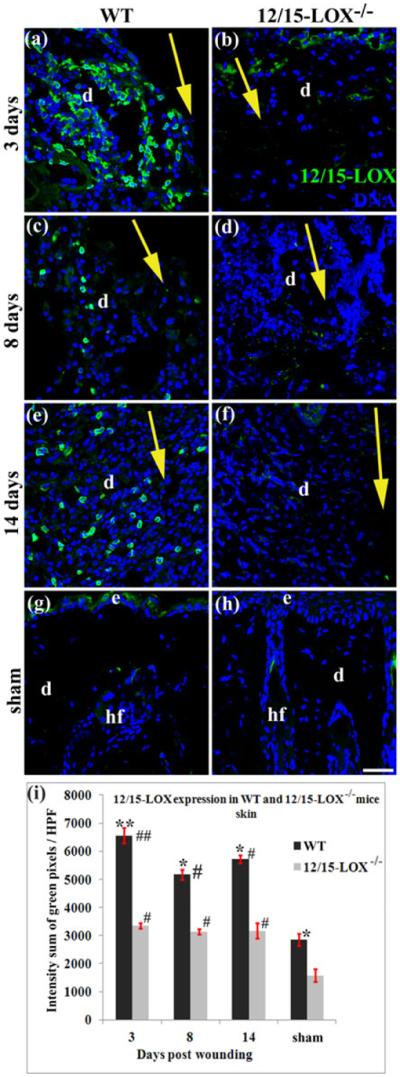
Fig. 6. 12/15-LOX expression in wounded and unwounded skin of 12/15-LOX−/− and WT mice.

12/15-LOX in wounded and unwounded skin were measured by an immunofluorescent histological method. Images in (a) and (b), images in (c) and (d), and image in (e) and (f), are for 3, 8 and 14 dpw, respectively. Images in (g) and (h) are for sham (unwounded skin). Images in (a), (c), (e) and (g), and images in (b), (d), (f) and (h), are for C57BL/6 (WT) and 12/15-LOX−/− mice, respectively. (i) Intensity summation of green fluorescent pixels per HPF of 12/15-LOX stained skin sections. Each value is the average of four mice per group ± SD. *p < 0.05 and **p < 0.01 are for WT versus 12/15-LOX−/−; #p < 0.05 and ##p < 0.01 are for the wounded versus sham of the same type of mice. Wound healing model or histological method was the same as in Fig 2. Nuclei are counterstained with DAPI (blue). HPF, high power field (40X); d, dermis; hf, hair follicle; Big yellow arrow, incisional wound and direction from epidermis to hypodermis of skin; Scale bar = 38 μm.
