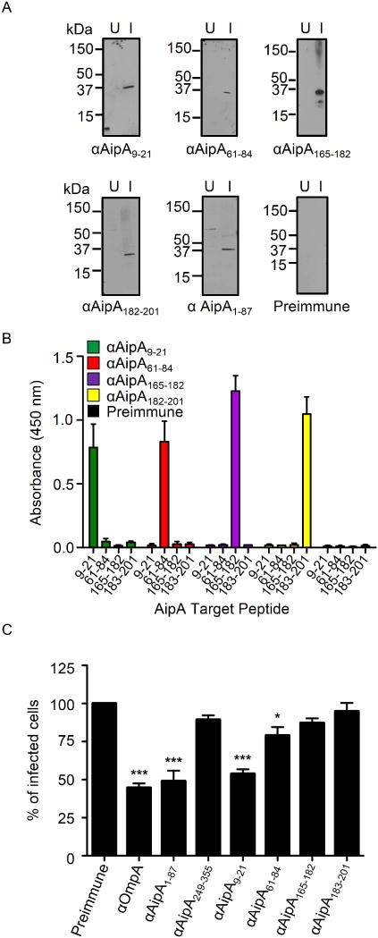
Figure 7. AipA residues 9–21 are critical for establishing infection in host cells.

(A) Western blot analyses in which rabbit antiserum targeting AipA9–21, AipA61–84, AipA165–182, AipA183–201, AipA1–87, or preimmune rabbit serum was used to screen whole cell lysates of uninfected (U) and A. phagocytophilum infected HL-60 cells (I). Data are representative of two experiments with similar results. (B) ELISA in which AipA9–21, AipA61–84, AipA165–182, and AipA183–201 antibodies were used to screen wells coated with peptides corresponding to AipA residues 9–21, 61–84, 165–182 and 183–201. Each antiserum only recognized the peptide against which it had been raised. Results shown are the mean (± SD) of triplicate samples. Data are representative of three experiments with similar results. (C) Pretreatment of A. phagocytophilum with AipA9–21 antiserum inhibits infection of HL-60 cells. DC bacteria were pretreated with antiserum specific for AipA9–21, AipA61–84, AipA165–182, AipA183–201, AipA1–87, AipA249–355, OmpA, or preimmune serum for 30 min. Next, the treated bacteria were incubated with HL-60 cells for 60 min. After removal of unbound bacteria, host cells were incubated for 24 h and subsequently examined using Msp2 (P44) antibody and confocal microscopy to assess the percentage of infected cells. Results shown are relative to preimmune serum-treated host cells and are the means ± SD for six experiments. Statistically significant (*, P < 0.05; **, P < 0.005; ***, P < 0.001) values are indicated.
