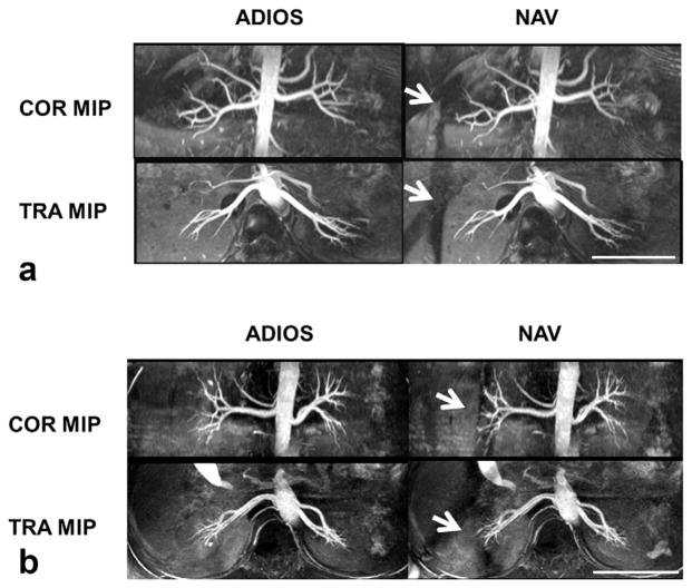
FIG. 3.
Representative coronal (COR) and transverse (TRA) maximal intensity projection (MIP) images from two healthy subjects comparing image quality of ADIOS and conventional diaphragm navigator gating (NAV), both acquired with spatial resolution of 1.1 × 1.1 × 2.2 mm3 and TR of two heartbeats. Scale bars represent 10 cm. Note the saturation bands caused by NAV (arrows in a,b). In some cases, NAV saturation bands degraded the visualization of distal right renal arteries (arrows in b), whereas in the ADIOS images there was no such effect.

