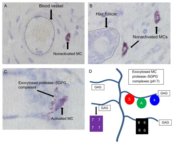
Figure 6.3.
Histochemistry of nonactivated and activated cutaneous MCs. Mature MCs are recognized in the skin and other tissues histochemically by the ability of their SGPGs to bind to toluidine blue and other cationic dyes. Cutaneous MCs tend to reside near blood vessels (A), hair follicles (B), and nerves (data not shown). When activated (C), cutaneous MCs quickly exocytose the content of their secretory granules which consist primarily of histamine and varied protease–SGPG complexes. Most of the cell’s positively charged granule proteases (D) (e.g., mMCP-5 (5), Cpa3 (A), mMCP-4 (4), and mMCP-6 (6)) are ionically bound so tightly to the negatively charged glycosaminoglycans (GAGs) of SGPGs that the exocytosed macromolecular complexes remain intact for hours in the ECM. An exception is mMCP-7 (7), due to its less positively charged SGPG-binding domain. The large size of these protease–SGPG complexes minimize their diffusion and ability to enter the circulation. Instead, most of them are eventually endocytosed by other cell types in the inflammatory site where they are destroyed in primary lysosomes. The depicted images in panels (A)–(C) were from surgically wounded mouse skin.

