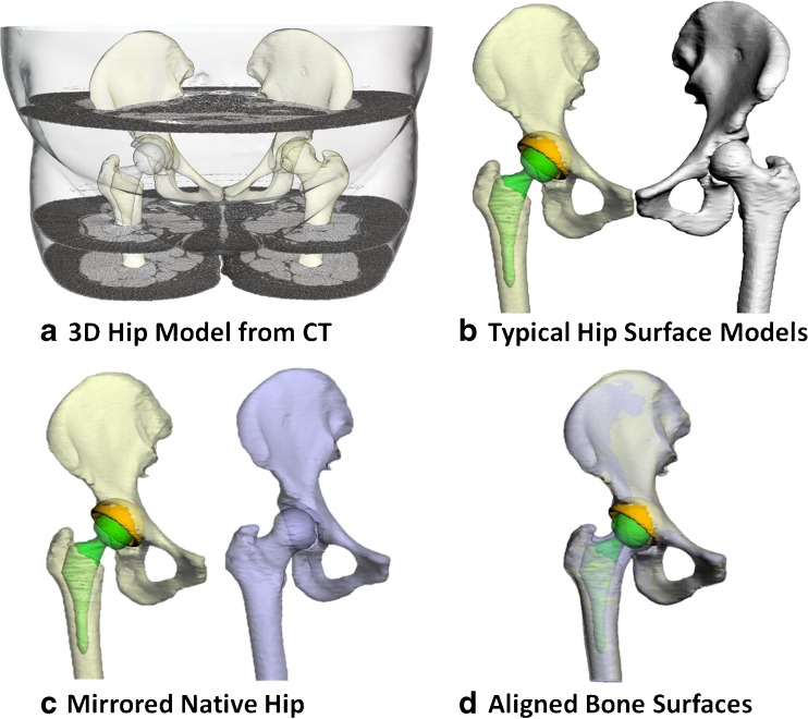
Fig. 1.
a Three-dimensional models of the hip bone, femur, acetabular cup and femoral stem reconstructed from computed tomography (CT) scan data. b Models were split into implanted and native groups. c The contralateral native hip model was mirrored with respect to the sagittal plane. d The mirrored hipbone and femur were then best aligned with the implanted hipbone and the remaining femur of the implanted side

