
Figure 9.
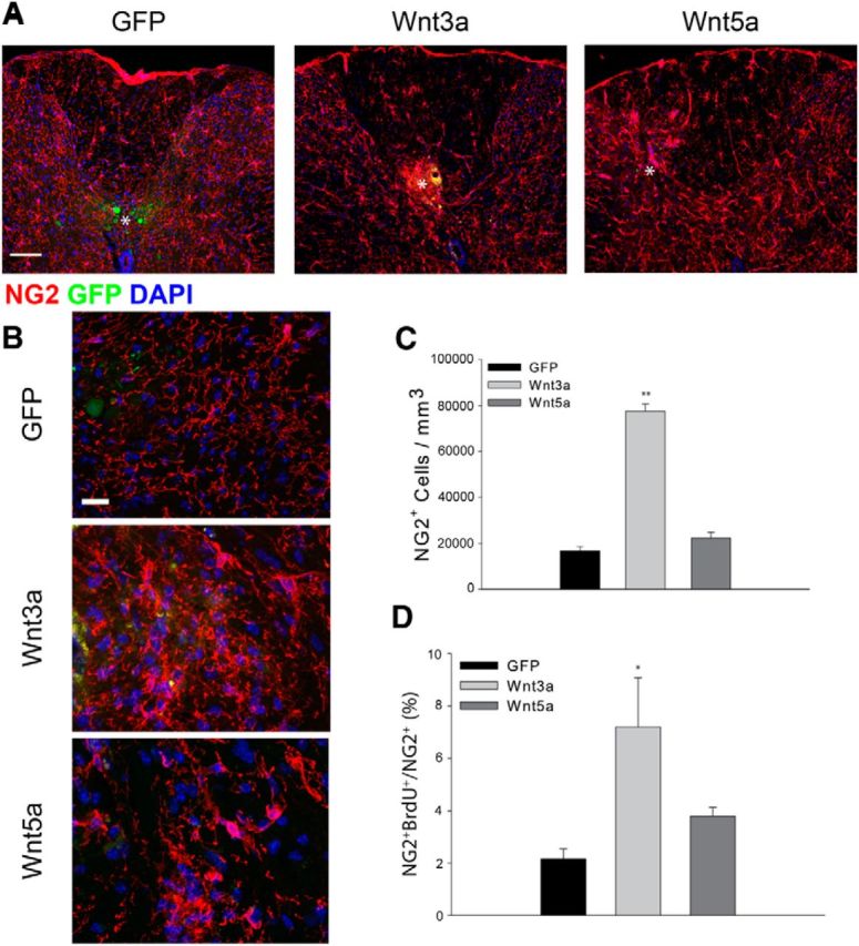
Exogenous Wnt3a increases OPC density. A, The density of NG2+ (red) OPCs increases in Wnt3a virus-injected animals but does not change significantly after injection of either a GFP virus or a Wnt5a-expressing virus. White asterisk indicates centroid of the injection site. B, High-power views of NG2 immunoreactivity after injection of the indicated AAV viruses. C, Quantitation of cell density 10 d post virus injection. D, BrdU incorporation into NG2+ cells after a pulse label. Wnt3a, n = 8; Wnt5a, n = 4; GFP, n = 6; **p = 0.001, *p = 0.01. Scale bars: A, 50 μm; B, 10 μm.