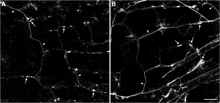
Figure 3.
Vagal afferent innervation pattern in the proximal small intestine. Low-magnification photomontages of vagal afferent innervation of the same region of the duodenum are shown in control (A) and INT-BDNF−/− (B) mice. These images illustrate the lattice pattern composed of circular and longitudinal axon bundles running in near-perpendicular orientations and the increased IGLE density and axon bundle diameter in INT-BDNF−/− mice compared with controls. Arrows denote IGLEs, and arrowheads indicate axon bundles. Scale bar: (in B) A, B, 2.5 μm.

