
Figure 2.
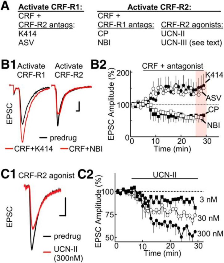
CRF-R1 activation potentiates EPSCs, and CRF-R2 attenuates ESPCs. A, Summary table listing the subtype selectivity for CRF receptor agonists and antagonists. The experimental conditions for selective activation of CRF-R1/R2 are described. B1, Stimulus-evoked EPSCs are increased by CRF-R1 activation (left) but decreased by CRF-R2 activation (right). To activate the CRF-R1 subtype, CRF (100 nm) was applied in the presence of K41498 (K414; 200 nm), a CRF-R2 antagonist. To activate the CRF-R2 subtype, CRF (100 nm) was applied in the presence of the CRF-R1 antagonist NBI27914 (NBI; 200 nm). Calibration: 100 pA, 1 ms. B2, Summarized time course of EPSC amplitude (% of baseline, mean ± SEM) illustrating the CRF-R1 potentiation and CRF-R2 attenuation with additional antagonists included for CRF-R1 (CP-154526, CP; 200 nm) and CRF-R2 (antisauvagine-30, ASV-30; 200 nm). Sample size for K41498, ASV, NBI, and CP154156 experiments: n = 6 cells at 3 or 4 rats per drug. C1, Sample stimulus-evoked EPSCs are attenuated by direct activation of CRF-R2 by UCN-II (300 nm). Calibration (all traces): 100 pA, 1 ms. C2, Summarized time course of EPSC amplitude (% of baseline, mean ± SEM) showing the concentration dependence of UCN-II (3–300 nm; sample size: n = 8 cells and 5 rats per concentration).