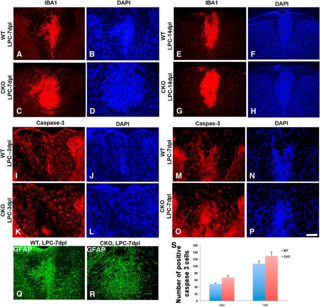
Figure 9.
Enhanced numbers of microglia and increased astrogliosis but modest increase in cell death detected in LPC-induced lesions in Cdk5 cKO mice compared with WT. Immunofluorescent staining of IBA1 (red) in LPC-lesioned Cdk5 cKO (C, D) and WT (A, B) mice at 7 dpl and in LPC-lesioned Cdk5 cKO (G, H) and WT (E, F) mice at 14 dpl. No significant difference in total number of immunostained activated Caspase-3 (red) between LPC-lesioned Cdk5 cKO (K, L) and WT mice (I, J) at 3 dpl and between LPC-lesioned Cdk5 cKO (O, P) and WT mice (M, N) at 7 dpl. DAPI (blue) was used as nuclei count staining. Increased level of astrogliosis marked by immunostained GFAP+ cells (green) in LPC-lesioned Cdk5 cKO (Q) and WT mice at 7 dpl (R). S, Quantification analysis showed the total number of activated caspase-3+ cells. Values are mean ± SEM. *p < 0.05, statistically significant. Scale bar, 25 μm.

