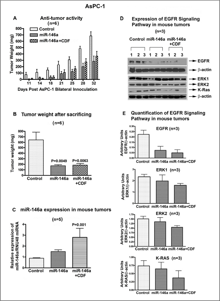
Figure 4.
Over-expression of miR-146a using stably transfected Pre-miR-146a followed by CDF treatment in AsPC-1 cells in vivo showed decrease in tumor growth rate (A). Tumor weight was significantly reduced in pre-146a and pre-146a+CDF, compared to control, in a xenograft model (B). This was consistent with re-expression of miR-146a in miR-146a and miR-146a+CDF group (C), which was associated with decreased EGFR, ERK1, ERK2, and K-Ras expression, compared to control (D). The relative expression of EGFR, ERK1, ERK2, and K-ras proteins were quantified against β-actin (E). P values relative to controls are mentioned over the bars

