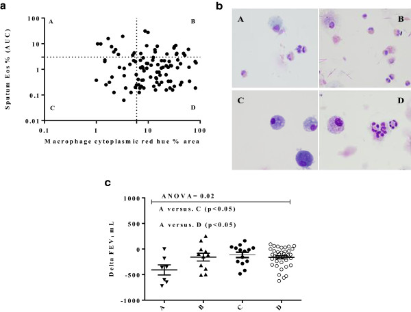
Figure 2.

Cytoplasmic macrophage red hue as an indirect measure of macrophage efferocytosis of eosinophils. a) Percentage area of sputum macrophage cytoplasmic red hue in COPD subjects against sputum eosinophil area under the curve (AUC) %/year. The cut-off points for the upper limit of the normal ranges are as shown on the horizontal and vertical axes, 6 (16) and 3% respectively. b) Representative images of sputum macrophages: A: Subjects with high eosinophils ≥ 3% and low red hue <6%; B: high eosinophils ≥3% and high red hue ≥6%; C: low eosinophils <3% and low red hue <6%; D: low eosinophils <3% and high red hue >6%. Group B and D subjects have purple coloured cytoplasm in their macrophages. Group A and C have light blue cytoplasm. c) Change of FEV1 between stable and first exacerbation visits for the 4 groups. The lines represent mean (SEM).
