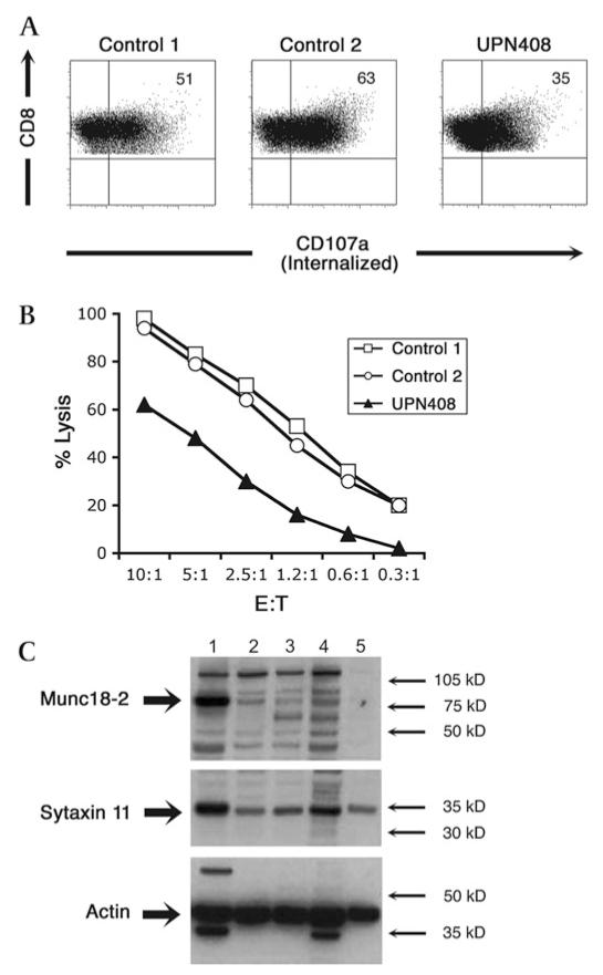
Figure 4. CTL degranulation and cytotoxicity and protein study of patient UPN 408.
Degranulation assay showing CD3 stimulated internalisation of CD107a (panel A), detected with PE-CD107a mAb present throughout the 4 h incubation, from CTL lines from two healthy donors (controls 1 and 2) compared to those from UPN408. Cells were stained for CD8 (y axis) and CD107a-PE (x axis). Cellular cytotoxicity of CTL lines from UPN408 (shaded triangles) compared to those from healthy donors (open symbols) measured using redirected lysis of P815 target cells at different E:T ratios (panel B). Data points were performed in triplicate with standard deviations of <2.5% for all E:T ratios. Western blot of cell lysates from CTL lines (1–4) or phytohaemagglutinin (PHA) blasts (5) from (1) Healthy donor, (2) FHL-5 patient with Pro477Leu mutation (see reference 24), (3–4) UPN408 clones, and (5) UPN408 PHA blasts probed with antibodies to Munc18-2, syntaxin 11 and reprobed with actin to show loading levels (panel C). Molecular weight markers are shown.

