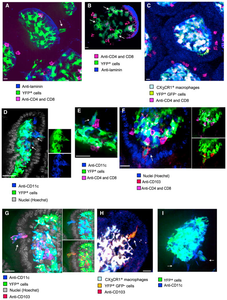
Figure 2. Immunohistology of the Epithelium Identifies CD11c-YFPint Cells as CD103+ DCs.
Samples of the small intestine from untreated mice were rapidly excised and fixed for immunohistological analysis of whole-mounted tissues. Single Z-planes are shown (scale bars represent 10 μM).
(A) In Cd11c-YFP mice, not only CD4+CD8+ IELs but also some CD11c-YFP+ cells (arrow) were located directly above the laminin+ basement membrane among enterocytes. Such cells expressed less YFP than the highly-branched cells confined to the LP.
(B) Within the epithelial layer, amoeboid YFP+ cells (arrows) could be seen alongside IELs.
(C) A CX3CR1-GFP−CD11c-YFP+ cell (in yellow) in the epithelium exhibited an elaborate shape and no T cell markers
(D) YFPint cells (arrows), located beneath and among enterocytes expressed membranal CD11c.
(E) A YFPint cell (arrow) expresses CD11c, but not CD4 or CD8, whereas adjacent IELs do.
(F) CD103 is expressed by the large YFP+ cells (arrow), which do not express CD4 or CD8.
(G) CD103 and CD11c are coexpressed by a large YFP+ cell (arrow) in the epithelium.
(H) CX3CR1−GFP−CD11c-YFPint cells, marked with membranal CD103+, can be located in the epithelium.
(I) YFP+ CD11c+ cells were also observed in the epithelium of Rag1−/− mice (arrows). All micrographs are representative of at least six independent samples.

