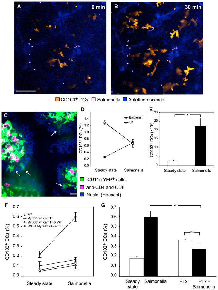
Figure 4. Luminal Salmonella Recruit CD103+ DCs to the Epithelium.
(A and B) Two-photon microscopy of the epithelial layer in several adjacent villi exposed to Salmonella. (A) Some intraepithelial YFPint CD103+ DCs were already visible at time 0, but within 30 min (B) they were joined by several other DCs that had migrated up into the epithelium (scale bar represents 50 μM).
(C) Immunohistology following Salmonella challenge captured several CD103+ DCs (arrows) within the epithelium. Image is representative of three experiments.
(D and E) By flow cytometry, Salmonella challenge increased the percentage of CD103+ DCs in the epithelium (p < 0.0001) and concomitantly decreased it in the LP (p = 0.003, data pooled from four experiments; percentages of DCs were calculated out of total live cells). (E) This translated to a marked increase in the total number of CD103+ DCs in the epithelium (p < 0.0001).
(F) We examined the ileal epithelium of WT mice, mice deficient in MyD88 and Ticam1 (TRIF) and chimeric mice whose hematopoietic cells are either MyD88−/− × Ticam1−/− or WT. Myd88−/− × Ticam1−/− mice had significantly lower percentages of intraepithelial CD103+ DCs (p = 0.012) and only in WT mice did Salmonella challenge significantly recruit CD103+ DCs, as indicated by ANOVA (p < 0.001, n = 27, data were pooled from two experiments and percentages calculated out of total live cells).
(G) Overnight treatment of mice with PTx significantly reduced the recruitment of CD103+ DCs to the epithelium (significant interaction at p < 0.001, n = 18, data pooled from two experiments). Error bars indicate SEM. Columns depict the percentages of CD103+ DCs out of total live cells.

