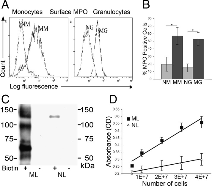
Fig. 2.
G-CSF up-regulates expression of catalytically active MPO on the surface of mobilized myeloid cells. (A) Representative MPO flow cytometry histograms are shown of native granulocytes (NGs) and native mononuclear cells (NMs) in comparison with mobilized granulocytes (MGs) and mobilized mononuclear cells (MMs). (B) Cumulative results of flow cytometry analysis for MPO cell surface expression typical for mononuclear cells and granulocytes. Values represent mean ± SD of percent positive cells from multiple donors (n = 15), *P < 0.001. (C) Representative Western blot of MPO immunoprecipitates (mAb 1A1) from lysates of surface biotinylated (+) or nonbiotinylated (−) MLs and NLs resolved on reducing SDS/PAGE gel. Biotin-labeled (membrane expressed) MPO was revealed with streptavidin-HRP. (D) MPO activity on the surface of NL and ML was evaluated by spectrophotometric detection of o-phenylenediamine dihydrochloride (OPD), a chromogenic peroxidase product (n = 5).

