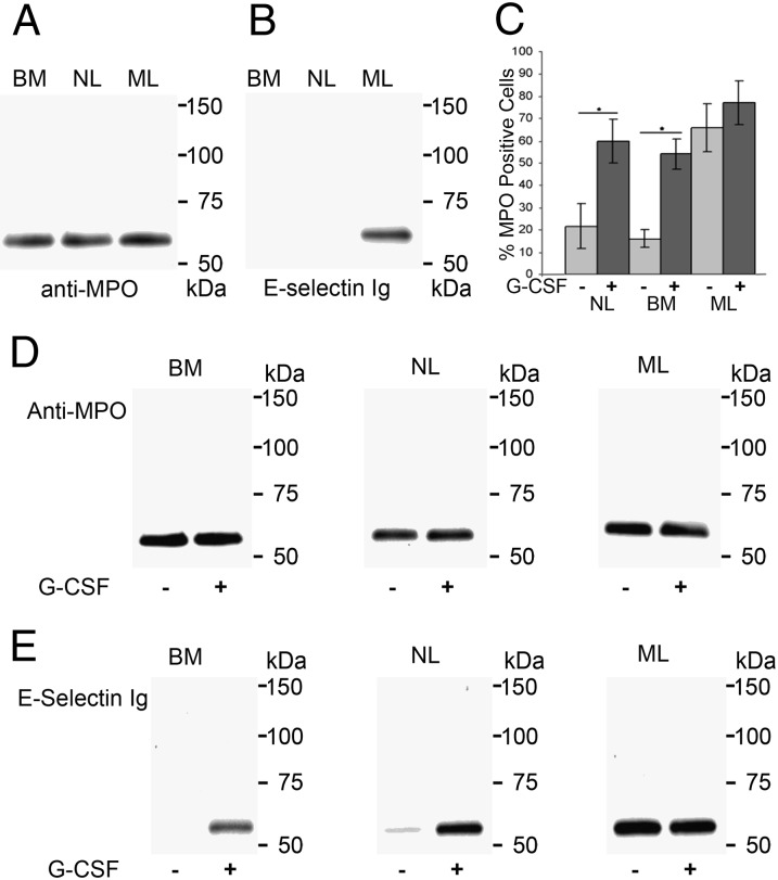
Fig. 3.
G-CSF induces cell surface expression and E-selectin ligand activity of MPO. (A) MPO immunoprecipitates (IPs) from lysates of bone marrow myeloid cells (BMs), NLs, and MLs stained in Western blot with anti-MPO mAb clone 3D3. (B) MPO IPs resolved on reducing SDS/PAGE and stained with E-selectin Ig chimera. (C) Flow cytometry analysis of NLs, BMs, and MLs cultured for 48 h without G-CSF (−) or with G-CSF (+). Values represent mean ± SD of percent MPO-positive cells from multiple donors (n > 30), *P < 0.001. (D) Western blots of MPO IPs from BMs, NLs, and MLs treated (+) or not treated (−) with G-CSF and stained with anti-MPO mAb 3D3. (E) MPO IPs from BMs, NLs, and MLs treated (+) or not treated (−) with G-CSF were probed with E-selectin–Ig chimera in Western blots.

