Fig. 3.
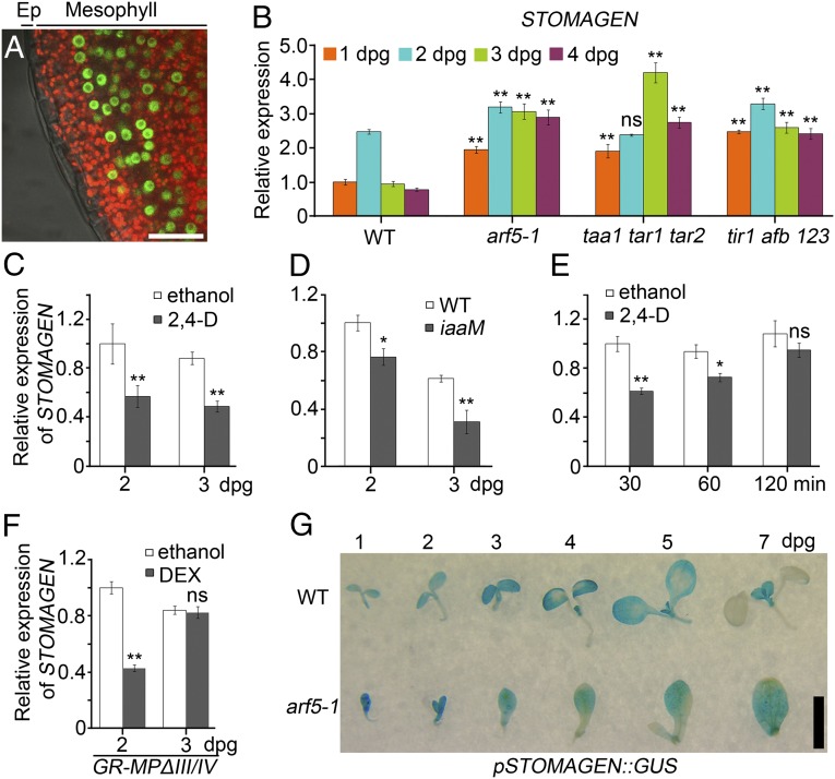
MP-mediated auxin signaling represses STOMAGEN expression. (A) Confocal images of pMP::MP-GFP leaves at 3 dpg. The merged signals of bright field, Cy5, and GFP channels are shown. Cy5 shows chlorophyll autofluorescence (red) in mesophyll. The nuclear GFP signals (green) indicate MP-GFP. Ep, epidermis. (B–F) STOMAGEN qRT-PCR in the seedlings with indicated genotypes and/or treatments. Data are means ± SDs (n = 3) with Student t test (*P < 0.01; **P < 0.001; ns, no significant differences). Statistical analyses are made by comparing data from mutants (B) and 35S::iaaM (D) with those from WT at the same age, respectively. (C) Seeds were grown in medium with ethanol control or 150 nM 2,4-D for indicated time, and statistical analyses are made by comparing data from different treatments at the same age. (E) The 2-d-old seedlings were treated in liquid medium with ethanol control or 150 nM 2,4-D for indicated time, and statistical analyses are made by comparing data from different treatments at the same time point. (F) 35S::GR-MPΔIII/IV seeds were grown in medium with ethanol control or 5 μM DEX for indicated time, and statistical analyses are made by comparing data from different treatments at the same age. (G) Time-course GUS staining of WT-like (WT) and arf5-1 siblings segregated from pSTOMAGEN::GUS;arf5-1/+. (Scale bars: A, 25 μm; G, 2.5 mm.)

