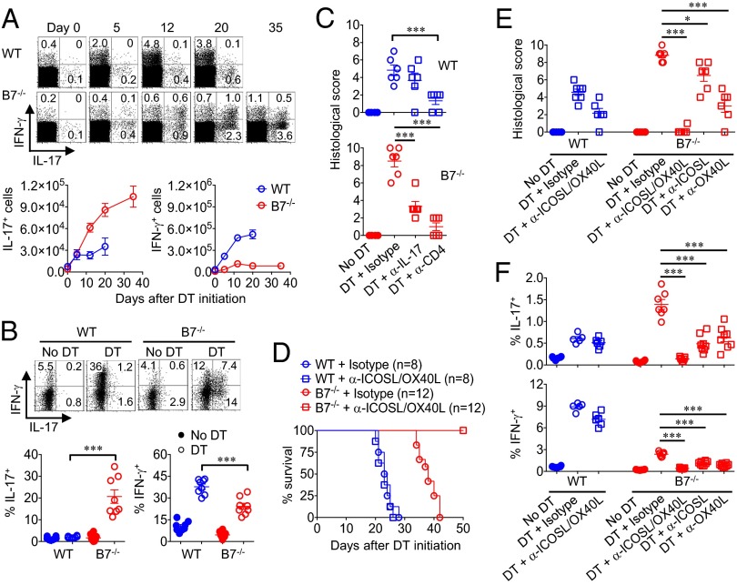
Fig. 3.
B7 deprivation drives expansion of Th17 CD4+ T cells through ICOSL/OX40L stimulation. (A) Percentage and number of IL-17– or IFN-γ–producing mesenteric lymph node CD4+ T cells in WT compared with B7-deficient Foxp3DTR mice at the indicated time points after initiating sustained Treg ablation. (B) Percentage of IL-17– or IFN-γ–producing lamina propria CD4+ T cells in WT compared with B7-deficient Foxp3DTR day 12 after initiating sustained Treg ablation. (C) Colonic inflammation disease score in WT (Upper) compared with B7-deficient Foxp3DTR (Lower) mice treated with either anti–IL-17A, anti-CD4, or isotype control antibody day 12 after concurrent DT administration for Treg ablation. (D) Survival for WT or B7-deficient Foxp3DTR mice after initiating sustained DT treatment and administration of anti-ICOSL/OX40L or isotype control antibodies. (E) Colonic inflammation disease score day 12 after initiating sustained Treg ablation for each group of mice described in D. (F) Percentage of IL-17– or IFN-γ–producing mesenteric lymph node CD4+ T cells day 12 after initiating sustained Treg ablation for each group of mice described in D. These data are representative of at least three independent experiments, each with similar results. Error bars indicate mean ± SEM. *P < 0.05, **P < 0.01, and ***P < 0.001.

