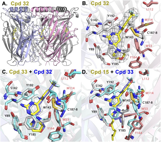
Fig. 2.
X-ray crystal structures of ligands 32 and 33 (negative cooperativity, nH < 1) and 15 (positive cooperativity, nH > 1), in complexes with Ls-AChBP. (A) Radial view of Ls-AChBP pentameric structure in complex with 32. Full occupation of the 10 binding sites in the unit crystal of a dimer of pentamers was evident. A principal, C loop-containing, and complementary face are shown in gray and purple. (B) Expanded radial view of 32 in binding site, including ligand electron density. Side chain carbons of the principal and complementary subunits are shown in gray and purple, respectively. Ligand carbons are in yellow and fluorines in turquoise. (C) Overlay of 32 (blue) and 33 (yellow) crystal structures. Side-chain carbons for 32 are in turquoise. The overlay shows little or no variance in ligand pose or side-chain positions. (D) Superimposition of 15 (yellow) and 33 (blue) crystal structures. Side-chain carbons for 33 are in turquoise. The positively 15 and negatively 32/33 cooperative ligands show a similar positioning of the 4-substituted phenyl rings, but distinct poses or positions for the 2-aminopyrimidine ring and the substituents at position 4 of the pyrimidine ring. Marked changes in the orientation of the side chains of Y185, W53, and Y164 are evident for the positively and negatively cooperative ligands.

