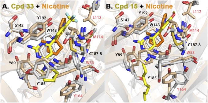
Fig. 3.
Superimposition of Ls-AChBP X-ray crystal structures in complex with 33 (A) and 15 (B) with nicotine (PDB code 1UW6) (in orange). 33 and 15 carbons are shown in yellow, and nicotine in orange. The protein side chains are shown in gray for 33 and 15 and pink for nicotine. Both ligands show distinctly different positions from the pyrrolidine and pyridine rings of nicotine, as well as the side-chain positions of residues in the C loop in the principal subunit (Y89 and Y185) and the complementary (W53 and Y164) subunit face.

